ACRAL FIBROKERATOMA
What is an acral fibrokeratoma?
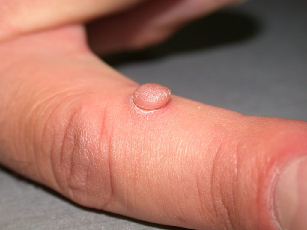
An acquired acral fibrokeratoma, also called digital fibrokeratoma, is an acquired growth that resembles a cutaneous horn located on the finger. The growth is a solitary, skin-colored, dome-shaped papule that is commonly less than 1.5cm in height or diameter, although some giant fibrokeratomas have been documented. An important finding to help differentiate these lesions from other look-alikes is the presence of a collarette of raised skin that encircles the base of the lesion, creating a moat. These lesions can also occur on the proximal hand, toes, soles or under the nail bed (periungual fibrokeratomas). Generally acral fibrokeratomas are benign lesions that are cosmetically bothersome. They have been reported in persons of all races and most commonly occur in middle aged persons with a slight male predominance.
How are acral fibrokeratomas formed?
The cause of acquired acral fibrokeratomas is unknown. Trauma has been implicated as a cause but no studies can confirm this hypothesis.
How are acral fibrokeratomas diagnosed?
Based on appearance acral fibrokeratomas can resemble other skin lesions, most commonly: corns, cutaneous horns, pyogenic granulomas, extra digits, warts or infantile acral fibrokeratoma (not acquired). The diagnosis of acral fibrokeratomas is one that must be made histologically by a pathologist. This is done by taking a small biopsy or sampling of the lesion and looking at it under the microscope.
How are acral fibrokeratomas treated?
Acral fibrokeratomas are benign lesions and do not require treatment unless cosmetically bothersome. Treatment is surgical requiring simple excision of the growth. After excision recurrence is rare.