ANGIOEDEMA
What is angioedema?
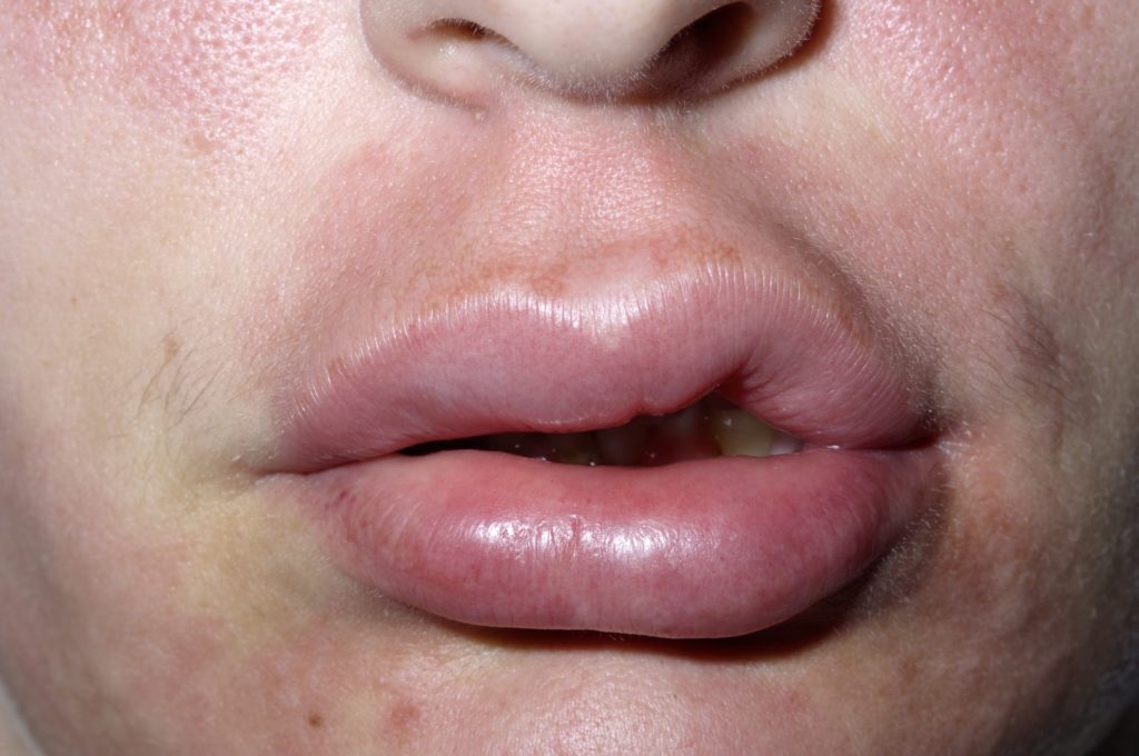
Angioedema is characterized by a large, swollen area that affects deeper layers of the skin (dermis and subcutaneous tissue). Because of its depth, angioedema is ill-defined and does not have sharp borders like those seen with hives (urticaria). The face (lips, eyes, earlobes, tongue), extremities and genitals are most commonly affected.
Urticaria and angioedema are caused by the same process but involve different depths of skin, they can occur together or independently. Angioedema is categorized into 2 groups: hereditary (without urticaria) and urticaria-associated (with urticaria). Patients with angiodema can present with itching and hypersensitivity over the swollen area. Angioedema may also be part of some syndromes, such as hereditary angioedema syndrome (HAE). One of the risks of angioedema is airway obstruction due to severe laryngeal edema.
What causes angioedema?
Angioedema is caused by an immune response involving specialized cells called mast cells. Mast cells release inflammatory proteins (histamine and bradykinins) that cause blood vessels to become leaky leading to deep tissue swelling. When angioedema occurs with urticaria it is usually due to an allergic reaction to an offending agent. HAE is caused by an enzyme deficiency that causes over-activation a pathway that leads to mast cell release. A certain class of blood-pressure lowering drugs called ACE-inhibitors may also trigger angioedema.
How is angioedema treated?
Clinical signs, history and skin testing can be used to make the diagnosis of angioedema. Once the diagnosis is made the initial goal of therapy is airway management. Systemic anti-histamines and epinephrine can be given to decrease the immune-response. Preventative management and avoiding the offending agent is important in urticaria-associated angioedema. HAE is a chronic disease and control, not cure is the goal of treatment. This is accomplished with steroids, anti-fibrinolytic (mechanism unknown) and transfusions of fresh frozen plasma to replace the deficient enzyme.
References:
Wolff K, Johnson, RA. Fitzpatrick’s Color Atlas and Synopsis of Clinical Dermatology. Sixth Edition. 2009.