BASAL CELL CARCINOMA
What is basal cell carcinoma?
Basal cell carcinoma (BCC) is the most common skin cancer in humans. They typically appear on sun-exposed skin, are locally invasive, slow growing and rarely metastasize. The skin lesions are usually asymptomatic but bleeding with minimal trauma may be the first symptom. There are 5 types of skin lesions:
- Nodular BCC: These are raised, bumps that are translucent and pearly. The color can vary from skin colored to reddish with telangiectasia. Telangiectasia is the presence of small spider-like blood vessels on the surface of the lesion. Nodular BCC is well defined and feel firm. Occasionally the nodules may have erosions or small pinpoints of melanin coloring.
- Ulcerating BCC: These are ulcerated lesions (no skin covering) with rolled borders. These lesions may appear translucent, pearly and smooth with telangiectasia.
- Sclerosing BCC: This starts as a small patch that looks like a healed scar. The borders are poorly defined, skin colored and may have peppery pigmentation. This form of BCC tends to extend deeper into the tissue so wider excision is required.
- Superficial multicentric BCC: These lesions appears as thin plaques that may be pink or red. They have a fine threadlike border and telangiectasia. This is the only form of BCC that may have scales.
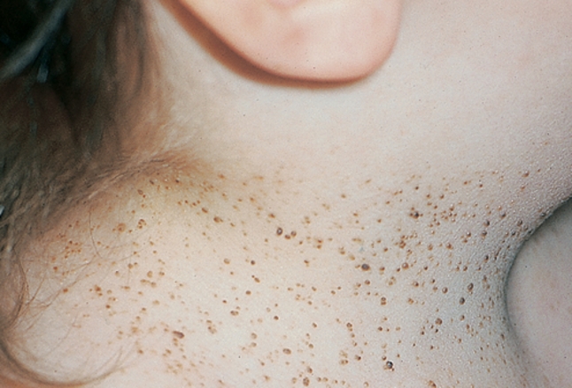
- Pigmented BCC: This lesion may be brown, blue or black and is smooth, hard and firm. It may look very similar to certain kinds of melanoma due to the pigment.
There are danger sites that are more likely to be affected by BCC, these include: inner and outer corners of the eyes, on either side of the nares, in the ear canal, on the scalp or behind ears.
What causes basal cell carcinoma and who is at risk?
Individuals with fair skin or those with high levels of UVB exposure are at increased risk for BCC. Previous therapy with x-rays (for facial acne) or arsenic exposure (even 30-40 years prior) increase the risk for BCC as well.
How is basal cell carcinoma diagnosed?
BCC is diagnosed by taking a biopsy and looking for certain characteristics under the microscope.
How is basal cell carcinoma treated and what is the prognosis?
Surgical excision with closure (primary, grafts or skin flaps) is the standard of therapy. Cryosurgery or electrocautery (cold or heat) are options but only for small lesions. Lesions on danger sites (see above) are best removed with Mohs surgery. Mohs surgery allows the surgeon to take a sampling of skin and view it under the microscope to ensure all of the tumor has been removed. Radiation therapy may be an option if surgical excision will cause disfigurement. There are a variety of topical treatments only for superficial BCCs below the neck.
References:
Wolff K, Johnson, RA. Fitzpatrick’s Color Atlas and Synopsis of Clinical Dermatology. Sixth Edition. 2009.