CAFÉ AU LAIT LESIONS
What are café au lait lesions?
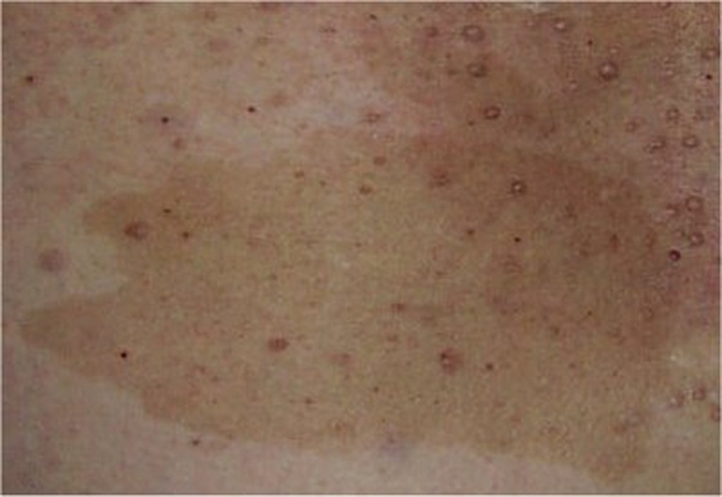
Café au lait (CAL) lesions are flat, pigmented spots (macules) that present on the skin within the first 3 years of life. These lesions are associated with neurofibromatosis 1 and 2 (NF1 and NF2). Neurofibromata (skin colored growths) appear during late adolescence in those affected with NF1. NF1 affects several organ systems, see neurofibromatosis for more information. CAL skin lesions have light to dark brown uniform melanin pigmentation with sharp borders. These lesions can vary in size from multiple “freckle-like” macules <2mm (see “axillary freckling”) to very large brown macules >20cm. The most common size is 2-5cm. CAL macules vary in number and can range from few to hundreds.
What causes café au lait lesions and who is at risk?
The exact cause of CAL lesions is unknown, but they are associated with NF1 and NF2. Diagnosis of NF can be made by looking at CAL lesions under a dermatoscope or with a Wood’s lamp. Using these devices special characteristics are seen such as melanin macroglobules. The presence of CAL alone does not make a diagnosis of NF unless patient has more than 6 lesions greater than 0.5cm in children (1.5cm in adults) AND axillary and inguinal CAL freckles. There are other diagnostic criteria for NF.
References:
Wolff K, Johnson, RA. Fitzpatrick’s Color Atlas and Synopsis of Clinical Dermatology. Sixth Edition. 2009.